Figure 3.
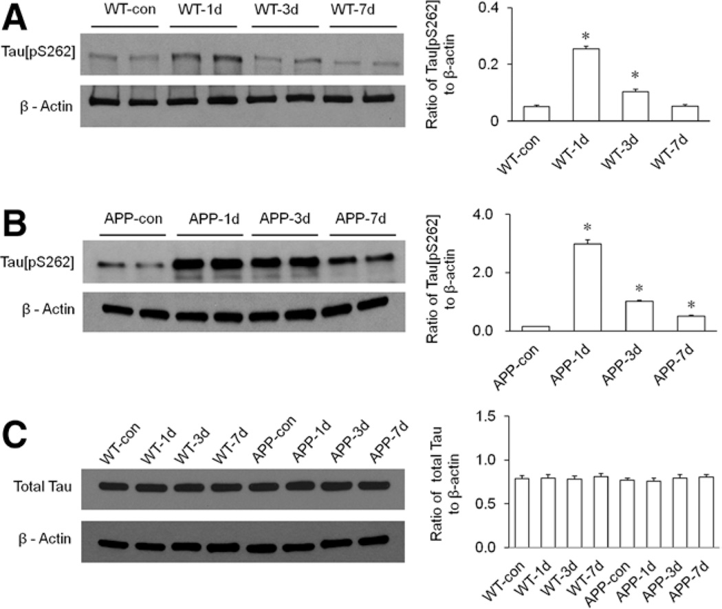
The effect of isoflurane exposure on hippocampal tau protein phosphorylation in both wild-type (WT) and transgenic amyloid precursor protein 695 (APP695) mice. All mice in isoflurane-treated groups received 1 minimum alveolar anesthetic concentration (MAC) of isoflurane for 4 hours. A, Isoflurane exposure time dependently altered the expression of tau[pS262] in the hippocampus of WT mice. Statistical analysis indicated that the levels of hippocampal tau[pS262] in WT mice were significantly increased on days 1 (WT-1d) and 3 (WT-3d) after isoflurane exposure (n = 6 per group, *P < 0.0001 for day 1 and *P = 0.0008 for day 3 versus WT-con group), and the tau phosphorylation returned to control level on day 7 (WT-7d) post-isoflurane (n = 6 per group, P = 0.8 versus WT-con group). B, Isoflurane exposure time dependently altered the expression of tau[pS262] in the hippocampus of the APP695 mice. Statistical analysis indicated that the levels of hippocampal tau[pS262] in the APP695 mice were significantly increased on days 1 (APP-1d), 3 (APP-3d), and 7 (APP-7d) after isoflurane exposure (n = 6 per group, *P < 0.0001 versus APP-con group). C, Isoflurane exposure had no effect on the expression of total tau protein in the hippocampi of both types of mice. Statistical analysis indicated that the levels of total tau protein in the hippocampi of all mice were not significantly different (n = 6 per group, WT: P = 0.95 for day 1, P = 0.87 for day 3, and P = 0.65 for day 7 versus WT-con group; APP695: P = 0.54 for day 1, P = 0.90 for day 3, and P = 0.75 for day 7 versus APP-con group). β-Actin served as a loading control for all Western blotting experiments.

