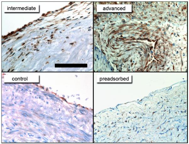
Figure 1. Immunostaining for carbamylated proteins in human atherosclerotic tissue.
Reddish-brown immunostaining indicates accumulation of carbamylated epitopes detected with an anti-HCit antibody mainly within or around the endothelial lining of human control tissue and intermediate atherosclerotic lesions. Intense staining was observed in all regions of advanced atherosclerotic lesions. Pre-incubation of the anti-HCit antibody with carbamylated albumin (pre-adsorbed) almost completely abolished staining. Scale bar represents 100μm.

