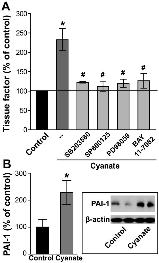
Figure 4. Increased expression of tissue factor and plasminogen activator inhibitor-1 (PAI-1) in cyanate-stimulated endothelial cells.
(A) Cyanate-treated HCAECs (1 mM, 4 h) show an increased expression of tissue factor, which is recovered in the presence of specific MAPK family member inhibitors (p38 MAPK inhibitor [SB203580; 5 μM] - JNK-2 inhibitor [SP600125, 5 μM] - ERK1/2 inhibitor [PD98059; 10 μM]) or the NF-kB inhibitor (BAY 11-7082; 5 μM). Tissue factor expression was determined by flow cytometry as described in Methods. Control was set at 100% and values are expressed as % of control. Results are shown as mean ± SEM (n= 4-5). Statistical analysis was performed by One-way ANOVA. Significance was accepted at * p <0.05 versus control, # p <0.05 versus cyanate-treated cells. (B) HCAECs showing enhanced PAI-1 expression after cyanate treatment (1 mM, 24 h). A representative Western blot is shown. Blots were analyzed with ImageJ software and normalized to β-actin. Data expressed as mean ± SEM (n=3). Student’s t-test was used for comparison. Significance was accepted at * p <0.05 versus control.

