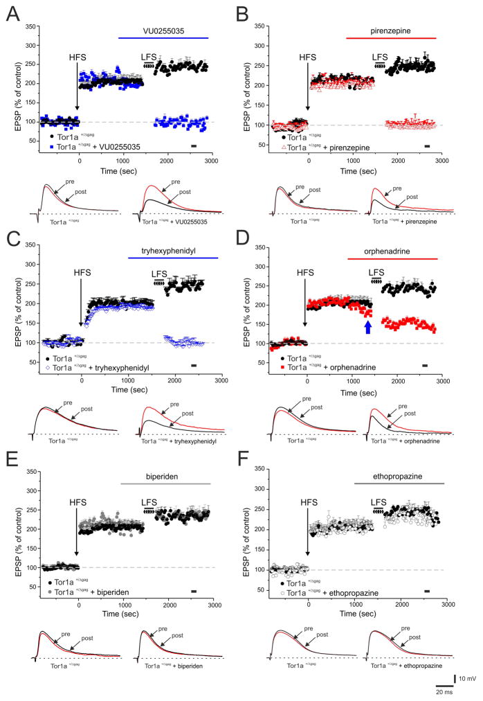
Figure 3. Antagonists of mAChRs restore synaptic depotentiation (SD) in Tor1a+/Δgag MSNs.
(A) After LTP induction, a low-frequency stimulation, LFS (2 Hz, 10 min) protocol fails to induce SD in Tor1a+/Δgag MSNs (black circles). Before LFS, bath-application of VU0255035 (100 nM, blue bar) restores SD in Tor1a+/Δgag mice. (B, C) Similarly, both pirenzepine (100 nM, red triangles) and tryhexyphenidyl (3 μM, blue diamonds), restore SD. Conversely, bath-applied orphenadrine (100 nM, red bar) causes only a partial rescue of SD in Tor1a+/Δgag MSNs (D) (blue arrow indicates the decrease in EPSP amplitude by orphenadrine). Biperiden (20 μM, grey filled circles) and ethopropazine (100 μM, grey open circles), fail to restore the LFS-induced SD (E, F). Below each plot representative EPSPs recorded before (pre) and 15 min after (post) LFS protocol. The black spot indicates at which time point samples were measured. Each data point represents the mean ± SEM of ≥8 independent observations.

