
FIG. 9.
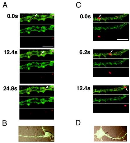
Axonal transport of hPVRα-GFP or hPVRMα-GFP-expressing vesicles in PC12 cells. PC12 cells transfected with hPVRα-GFP (A and B) or hPVRMα-GFP (C and D) were incubated with tetramethylrhodamine-conjugated dextran and PV and subjected to video rate scanning under a confocal microscope. The boxed areas in panels B and D were magnified and observed. Red shows the localization of dextran, and green shows the localization of hPVRs. The uppermost images at each time point are merged. The arrows indicate the vesicles containing dextran and hPVRs. Bars, 5 (A and C) and 10 (B and D) μm.