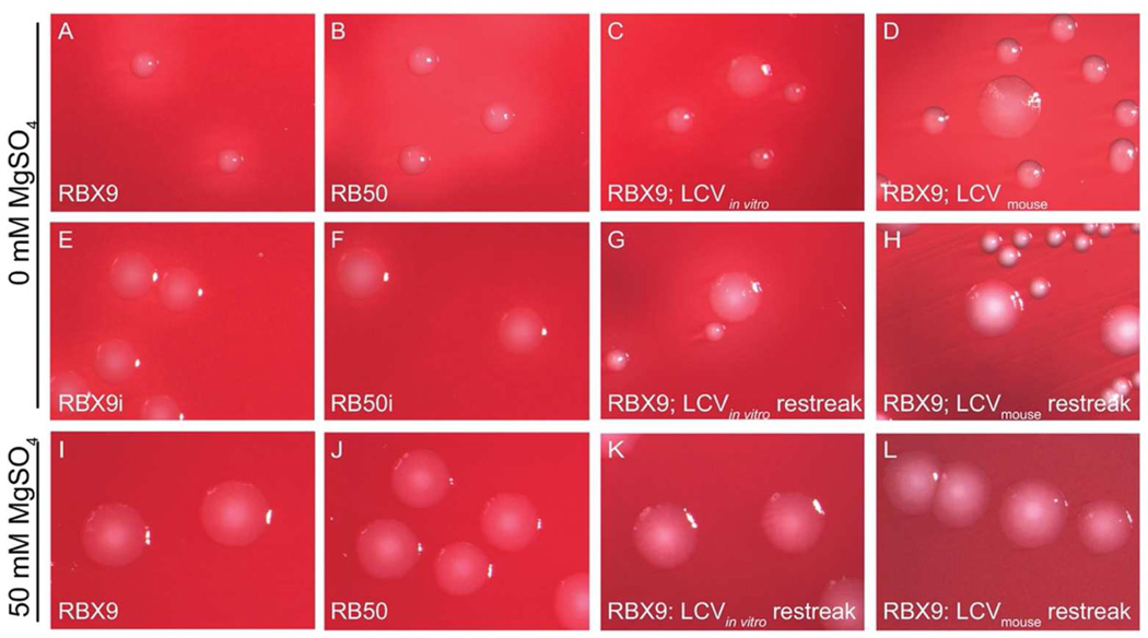
Figure 2.
RB50 and RBX9 colony morphology. Bacteria were plated on either BG agar or BG agar + 50mM MgSO4 and were imaged after 48h. A, RB50; B, RBX9; C, RBX9 LCV produced after in vitro modulation: D, RBX9 LCV recovered from mouse lung homogenate; E, RB50i (a Bvg-intermediate phase-locked strain in the RB50 background); F, RBX9i (a Bvg-intermediate phase-locked strain in the RBX9 background); G, RBX9 restreak of an LCV produced after modulation; H, RBX9 restreak of an LCV recovered from the mouse lung; I, RB50; J, RBX9; K, RBX9 restreak of an LCV produced after modulation; L, RBX9 restreak of an LCV recovered from the mouse lung.

