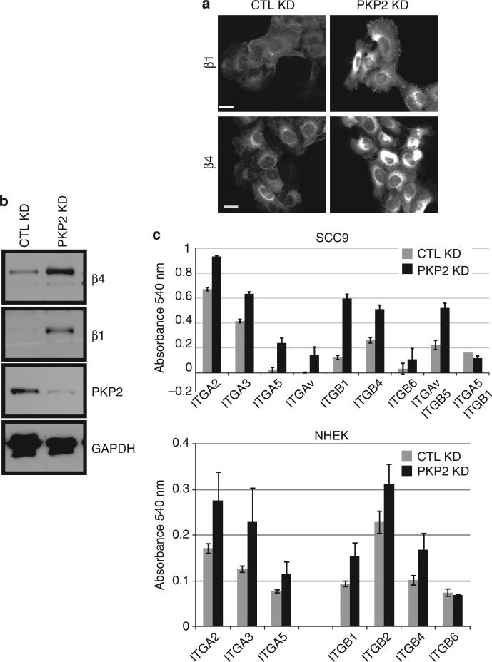
Figure 5. The expression of β1 and β4 integrin is increased upon plakophilin 2 (PKP2) silencing.
(a) Staining of control (CTL) and PKP2-silenced normal human epidermal keratinocytes (NHEKs) demonstrates an increased amount of β1 and β4 integrin expressed by the PKP2 knockdown (KD) cells. Scale bar = 20 μm. (b) Whole-cell lysates of NHEKs demonstrate an elevated amount of both β1 and β4 integrin in steady-state PKP2 KD keratinocyte cultures compared with control cells. GAPDH, glyceraldehyde-3-phosphate dehydrogenase. (c) An α and β integrin ELISA demonstrates an increased amount of α and β integrin subunits expressed on the surface of SCC9 cells or NHEKs silenced for PKP2 (error bars ±SEM).

