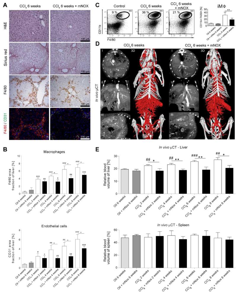
Figure 5. Effect of pharmacological inhibition of CCL2-dependent inflammatory monocytes on fibrosis-associated angiogenesis.
Chronic toxic liver injury was induced by repetitive i.p. administrations of CCl4 in c57BL/6 mice, and half of these animals received thrice weekly s.c. injections of the specific CCL2 inhibitor mNOX-E36, to block the CCL2-dependent infiltration of inflammatory monocytes. Analyses were performed 48 hours after the last CCl4 injection. Control mice received corn oil for 6 weeks. (A) Representative H&E staining, Sirius red, F4/80 immunohistochemistry, and F4/80-CD31 immunofluorescence co-stainings. (B) Quantification of F4/80+ macrophages and CD31+ blood vessels in livers of chronically injured and mNOX-E36 treated mice. (C) Representative FACS plots and statistical analysis showing the increase of intrahepatic inflammatory macrophages (iMΦ) in chronically injured livers and their significant reduction in mNOX-E36 treated livers. iMΦ were separated from Kupffer cells on the basis of differential expression of F4/80 and CD11b. (D) Liver vascularization visualized by contrast-enhanced in vivo μCT. (E) Quantification of the rBV in livers and spleens using functional in vivo μCT imaging. Data are shown as mean±SD. ***P<0.001, **P<0.01 and *P<0.05 for comparing CCl4 vs. CCl4+mNOX-E36; ###P<0.001, ##P<0.01 and #P<0.05 for comparing CCl4 or CCl4+mNOX-E36 vs. corresponding control groups (i.e. 6 weeks oil or 6 weeks oil+mNOX-E36) (n=30 mice; Student’s t test).

