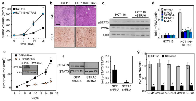
Figure 5. Colon tumor development in vivo critically depends on expression of STRA6.
a) NCr athymic male mice were injected with 2 x10⁶ HCT116 cells stably transfected with an e.v. into the right flank and HCT116 cells stably expressing STRA6 into the left flank. Tumor growth at both injection sites was monitored by measuring the length and width with calipers and tumor volume calculated as length x width2/2. Data are mean±S.E.M. (n=5)*p<0.05 vs. e.v.-expressing tumors. b) H&E staining and Ki67 immunostaining of tumors that arose from HCT116 cells expressing e.v or STRA6 (bar: 100 μm). c) Immunoblots of pSTAT3 and the proliferation marker PCNA in tumors that arose from HCT116 cells expressing e.v or STRA6. d) Levels of denoted mRNAs in tumors that arose from HCT116 cells expressing e.v or HCT116 cells expressing STRA6. Data are mean±S.E.M. (n=3)*p<0.05 vs. e.v.-expressing tumors. e) NCr male mice were injected with 5×10⁶ SW480 cells stably expressing either GFP shRNA (shGFP, left flank) or STRA6 shRNA (shS6, right flank). Tumor growth at both injection sites was monitored. Data are mean±S.E.M. (n=9). *p<0.01 vs. tumors expressing GFP shRNA. Inset: immunoblots of STRA6 in SW480 lines stably expressing GFP shRNA (shGFP) or STRA6 shRNA (shS6). f) Left: immunoblots of pSTAT3 in tumors that arose from SW480 cells stably expressing GFP shRNA or STRA6 shRNA. Right: quantification of immunoblots. Mean±S.E.M. (n=3). * p<0.05 vs. GFP shRNA-expressing tumors. g) Levels of mRNA for STAT3 target genes in tumors that arose from SW480 cells expressing GFP shRNA or STRA6 shRNA. Data are mean±SD (n=3). * p<0.01 vs. GFP shRNA-expressing tumors. All data are mean±S.E.M. (n=3).

