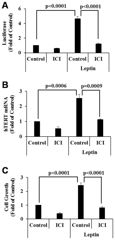
Figure 2. Leptin increases hTERT expression and cell growth through ERα activation.

A, BG-1 cells in 6-well plates were transfected with 0.5 μg pLEN-hERα, 0.5 μg EREe1bLuc and 0.2 μg pCMVβGal and 24 h later, treated for 72 h with vehicle (Control), leptin (100 ng/ml), 10−8 M ICI 182,780 (ICI) or leptin plus ICI as indicated. Luciferase activities were determined, normalized with β-gal activity and presented as fold of the vehicle control. B, BG-1 cells were treated as in panel A. Total RNAs were extracted and subjected to qRT-PCR analyses. The levels of hTERT were normalized with corresponding GAPDH and expressed as fold of the vehicle control. C, BG-1 cells were treated as in panel A but for 6 days. Cell growth was measured in MTT assays. Data represent three independent experiments. Error bars are SD. Statistical analyses were performed with Student’s t test (n=3 for panels A and B, n=10 for panel C).
