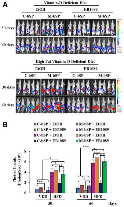
Figure 5. EB1089 suppresses HFD-induced ovarian tumor growth in vivo through miR-498.
Luciferase-marked BG-1 cells (5×10⁶) expressing control (C-bSP) or miR-498 (M-bSP) sponges were mixed with Matrigel and i.p injected into female athymic nu/nu mice fed with VDD or HFD. Mice bearing BG-1 xenograft tumors were treated by gavage every other day with EB1089 (0.5 μg/kg) or a vehicle (EtOH) in sesame oil. A, After 30 and 60 days of treatments, tumor growth was monitored by bioluminescent in vivo imaging. B, The bioluminescence intensity, a measurement of viable tumor volume, was quantified and presented as photon counts (photons/sec/cm2/steradian). Statistical analyses were performed with Student’s t test (n=5). #p<0.01; *p=0.002; **p=0.001; ***p<0.0001.

