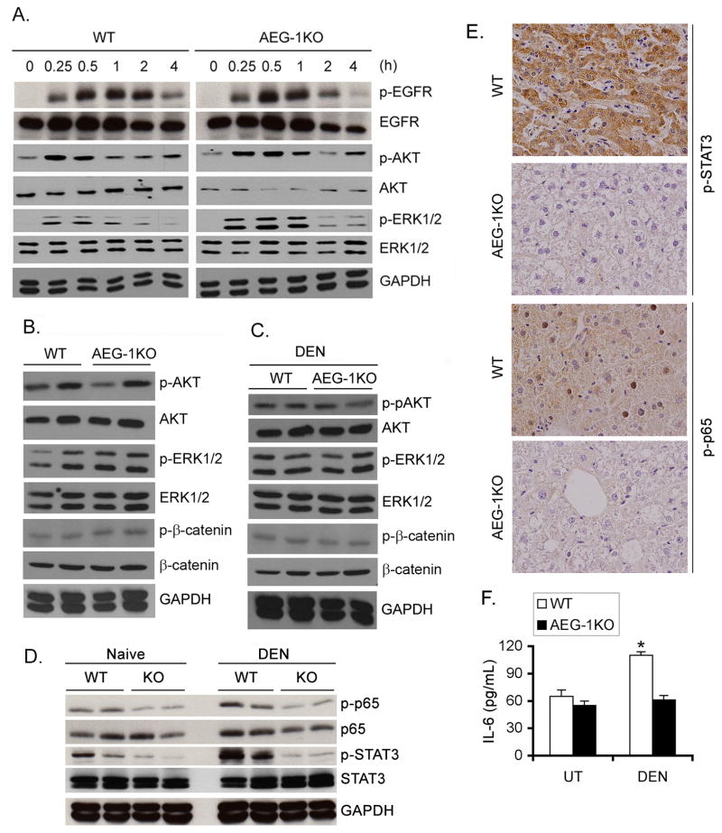
Fig. 3.
NF-κB and STAT3 activation is inhibited in AEG-1KO mouse. A. WT and AEG-1KO hepatocytes were treated with EGF (50 ng/mL) for the indicated time points and Western blotting was performed for the indicated proteins. B. Western blot was performed for the indicated proteins using liver lysates from adult WT and AEG-1KO mice. C. Western blotting was performed for the indicated proteins using liver lysates from DEN-treated WT and AEG-1KO mice at the end of the study. D. Western blotting was performed for the indicated proteins using liver lysates from naïve and DEN-treated WT and AEG-1KO mice at the end of the study. For B–D, each lane represents one independent mouse. E. DEN-treated liver sections were stained for p-STAT3 and p-p65 NF-κB at the end of the study. F. IL-6 protein level was measured in DEN-treated liver homogenates by ELISA. Data represent mean ± SEM. n = 5. *: p<0.01.

