
FIG. 1.
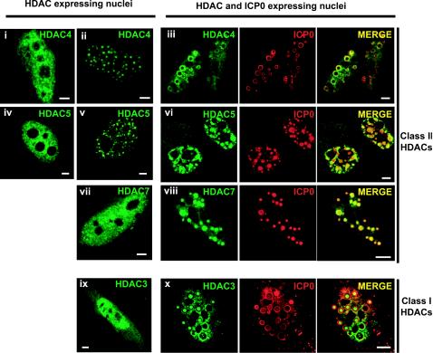
Nuclear redistribution of ectopically expressed epitope-tagged class I or II HDACs in HeLa cells expressing ICP0. Nuclear distribution of HA epitope-tagged class II HDAC4 (i to iii), 5 (iv to vi), 7 (vii and viii), and FLAG epitope-tagged class I HDAC3 (ix and x) expressed either alone (left images) or with ICP0 (right images). ICP0 and tagged HDACs were detected by using anti-ICP0 rabbit polyclonal antibody R190 and anti-HA (12CA5) or anti-FLAG (M5) MAbs. The secondary antibodies used were Alexa 488-conjugated goat anti-mouse (1/200) and Cy5-conjugated goat anti-rabbit (1/200) antibodies. Cell samples were examined with a Zeiss LSM 510 META confocal microscope with two lasers giving excitation lines at 488 and 633 nm as described in Materials and Methods. Bars, 5 μm.