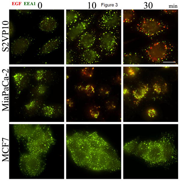
Figure 3.
Uptake of EGF-750 Probe detected via Immunocytochemistry. To demonstrate retention of biopotency of EGF in a validated fluorescent probe complex, Texas Red-EGF was incubated with three pancreatic cancer cell lines and analyzed at various time points (0, 10, and 30 min). S2VP10 and MiaPaCa-2 cell lines show internalization of labeled EGFR-Texas Red-EGF complex (red), while MCF7 (low EGFR-expressing) cells exhibit significantly reduced receptor-ligand internalization even with similar levels of endocytosis activity, apparent via labeled EEA1 antibody (green).

