
Figure 4.
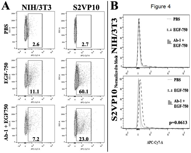
Extracellular accumulation of EGF-750 probe was assessed using flow cytometry. A, percentages represent proportion of APC-Cy7-positive tumor cells within gated area; values are representative of the mean of two experimental replicates. B, Wilcoxon signed-rank test determined differences in MFI was not significant among treatments (p=0.0613).