
FIG. 3.
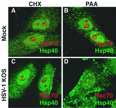
Analysis of chaperone localization in infected or uninfected cells treated with CHX or PAA. Merged images of cells stained with antibodies specific to Hsc70 (red) and Hsp40 (green) are shown. (A and B) Uninfected cells treated with CHX or PAA. (C and D) HSV-1-infected cells treated with CHX or PAA.