
FIG. 6.
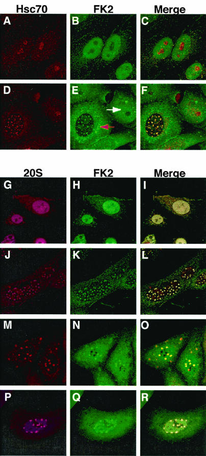
Localization of ubiquitinated proteins and components of the proteasomal machinery during HSV-1 infection. Uninfected (A to C) and HSV-1-infected (D to F) cells stained with antibodies specific to Hsc70 (A and D) and ubiquitinated proteins (FK2) (B and E) are shown. Merged images are shown in panels C and F. In panel E, the white arrow indicates the diffuse nuclear staining of ubiquitinated proteins in an uninfected cell and the red arrow indicates the dramatic redistribution of ubiquitinated proteins in an HSV-1-infected cell. The subcellular localization of the 20S core component of the 26S proteasome (G, J, M, and P) and of ubiquitin-conjugated proteins (H, K, N, and Q) is also shown. Merged images are shown in panels I, L, O, and R. (G to I) Mock-infected cells stained with α20S and αFK2 antibodies. (J to L) HSV-1-infected cells stained with the same antibodies. Infected cells treated with the proteasomal inhibitor MG132 throughout infection (M to O) or at 2 h postinfection (P to R) are also shown.