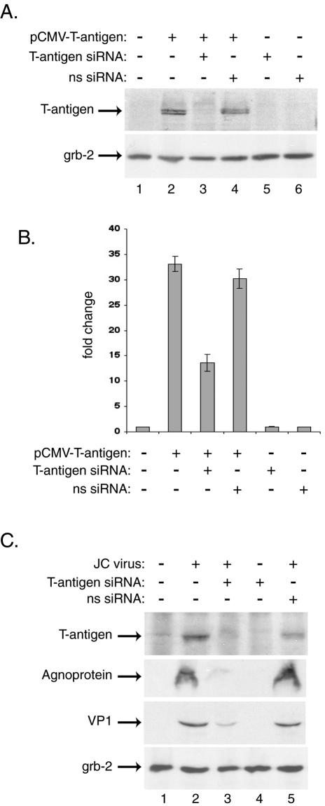
FIG. 1.

JCV T-antigen siRNA decreases expression of JCV proteins in transiently transfected and infected primary human astrocytes. Primary human fetal astrocytes were prepared as described previously and were seeded into six-well plates at a density of 500,000 cells/well (20). For transient transfections, cells were transfected by using FuGENE 6 with plasmid expressing JCV T antigen (15). The following day, the cells were transfected with double-stranded 21-bp siRNA for JCV T antigen targeting nt 4256 to 4276 of the Mad-1 isolate of JCV (sense strand, 5′-AAGUCUUUAGGGUCUUCUACCUdTdT-3′), while a nonspecific RNA (ns siRNA) targeted nt 4406 to 4426 of the reference strain 776 of SV40 (sense strand, 5′AAGUCCUUGGGGUCUUCUACCUdTdT-3′). The two base pair mismatches between the JCV and SV40 T antigens are underlined. The siRNAs were prepared as double-stranded, 2′-deprotected, and desalted oligonucleotides and were utilized according to the manufacturer's directions (Dharmacon). For the transfection of siRNAs, 100 pmol of siRNA was mixed with 3 μl of Oligofectamine (Invitrogen), diluted in OptiMEM (Invitrogen), and incubated with the cell cultures for 4 h at 37°C under serum- and antibiotic-free conditions. After transfection, the cells were fed with serum-containing medium without removing the siRNA transfection mixture. (A) Whole-cell extracts prepared from transfected astrocytes 24 h after siRNA treatment were analyzed by Western blotting for the presence of T antigen (pAb416; Oncogene Science) and the unrelated protein Grb-2 (upper and lower panels, respectively). (B) In parallel, samples transfected with 1.0 μg of JCV T-antigen expression plasmid along with 0.5 μg of a luciferase reporter construct containing the JCV late promoter (Mad-1 strain) were harvested 24 h after siRNA treatment, and luciferase activity was measured according to the manufacturer's directions (Promega luciferase assay system). Activities are presented as fold changes from the background activity of the JCV late promoter, arbitrarily set as 1. Data are means from four experiments, and standard deviations are indicated by error bars. (C) Primary astrocytes were infected with the JCV Mad-4 strain at a multiplicity of infection of 1 in serum-free medium for 3 h at 37°C. Uninfected and infected cells were then transfected with T-antigen siRNA at days 1, 5, and 10 postinfection and were harvested at day 15. Western blotting was performed on whole-cell extracts for the presence of JCV early and late proteins T antigen (pAb416; Oncogene Research Products), agnoprotein (7), and VP1 (pAb597; kindly provided by Walter Atwood, Brown University) as well as the cellular protein Grb-2 (pAb81; BD Biosciences). Proteins were visualized by using horseradish peroxidase-conjugated secondary antibodies and the ECL-Plus system (Amersham).
