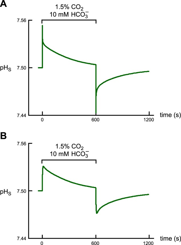
Fig. 3.

Effect of the new experimental protocol on the shape of the pHS trajectory and on the time to peak. A: pHS trajectory predicted by the model when using the protocol of Somersalo et al. (22). In using this original protocol, we assume that, at the outset of the simulation, the CO2/HCO3− is already at the plasma membrane. B: pHS trajectory predicted by the model when using the revised protocol. Here we assume that the oocyte is initially superfused with the CO2/HCO3−-free ND96 solution until we switch the bulk solution to one containing CO2/HCO3− and allow the CO2 and HCO3− to diffuse to the cell surface.
