Fig. 1.
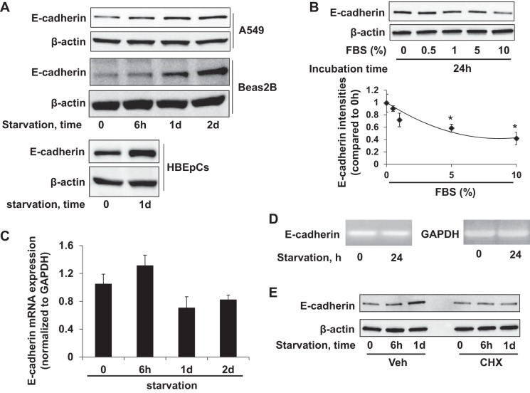
Serum starvation upregulates E-cadherin expression at the translational level. A: A549 or Beas2B cells were cultured in serum-free basal RPMI-1640 medium for 0, 6 h, 1 day (1d), and 2 days (2d). Human bronchial epithelial primary cells (HBEpCs) were cultured in growth factor-free medium for 1 day. E-cadherin and β-actin were detected by immunoblotting. B: A549 cells cultured with a range of serum concentrations (0, 0.5%, 1%, 5%, and 10%) for 24 h were analyzed for E-cadherin and β-actin by immunoblotting. Bottom: E-cadherin densitometries were normalized to β-actin intensities. *P < 0.05 compared with 0% of FBS. C: E-cadherin mRNA expression levels in A549 cells in serum-free media (0 h, 6 h, 1 day, 2 day) analyzed by qRT-PCR relative to GAPDH mRNA as internal control; fold changes were calculated relative to 0-h data. D: agarose gel image of E-cadherin and GAPDH full-length cDNA amplified by PCR with 28 cycles. E: cell lysates from serum-starved A549 cells treated with vehicle or 20 μg/ml cycloheximide (CHX) were analyzed by E-cadherin and β-actin immunoblotting. Blots shown are representative from 3 independent experiments except the bottom panel in A (2 experiments).

