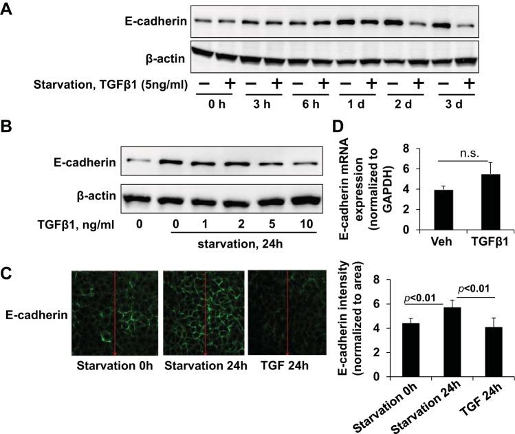
Fig. 4.
TGF-β1 attenuates serum starvation-mediated E-cadherin upregulation. A: A549 cells were cultured in serum-free basal RPMI-1640 medium for 0, 3 h, 6 h, 1 day, 2 days, and 3 days in the presence or absence of TGF-β1 (5 ng/ml) and lysates analyzed for E-cadherin. B: A549 cells treated with a dose range of TGF-β1 (0, 1, 2, 5, 10 ng/ml) in serum-free conditions for 24 h were analyzed for E-cadherin. C: A549 cells were serum-starved for 24 h in the presence of TGF-β1 (5 ng/ml), and E-cadherin (green) was visualized by immunocytochemistry under identical exposure conditions. Fluorescence intensity profiles shown at right. Immunoblots and images shown are representative from 3 independent experiments. D: E-cadherin mRNA level in TGF-β1 (5 ng/ml, 24 h)-treated A549 cells by qRT-PCR shown as fold changes relative to GAPDH control. ns, not significant.

