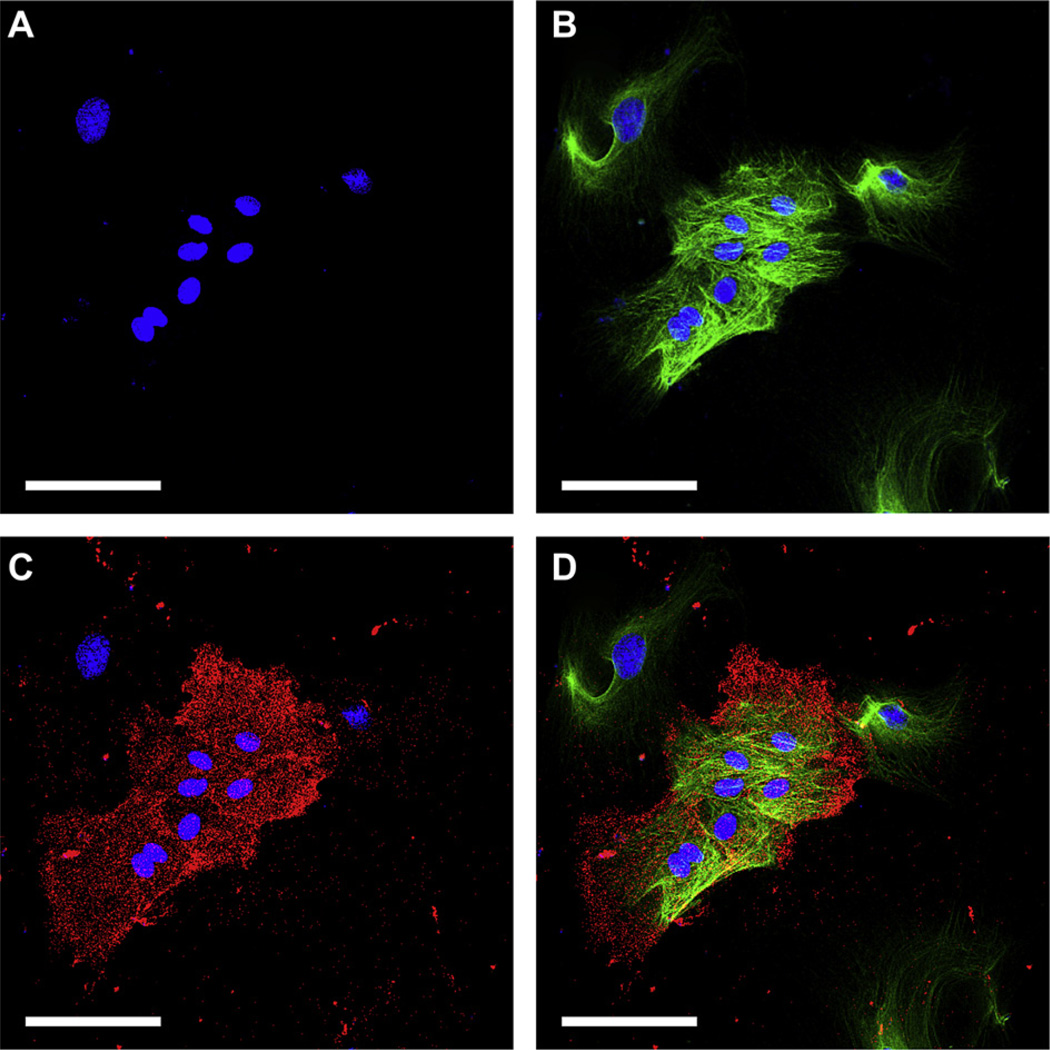
Fig. 5.
Laser confocal microscopy images obtained after 14 days of rat conjunctival primary culture showing co-expression of the putative stem cell marker ABCG2 and goblet cell marker CK-7. Nuclei were stained with DAPI (A, blue) and cells immunostained with anti-CK-7 (B, green) and ABCG2 (C, red). All cells are CK-7+ with a group of smaller cells co-expressing ABCG2 (D). Magnification: ×400. Scale bars: 100 µm.

