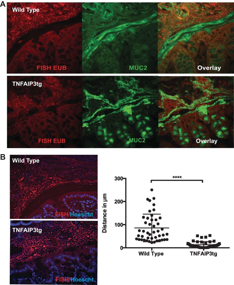
Fig. 6.
Microbial invasion of the intestinal inner mucus layer in villin-TNFAIP3 transgenic mice. A: fluorescence in situ hybridization (FISH) with EUB338 probe for bacterial 16S rRNA (EUB) (red) and immunohistochemistry for Muc-2 (green) in Carnoy's-fixed paraffin-embedded colonic tissue. B: FISH and Hoescht (blue) staining to detect the spatial separation of bacteria from epithelial cells with quantification of the distance (μM) between the epithelial surface and the bacterial population. ****P < 0.001.

