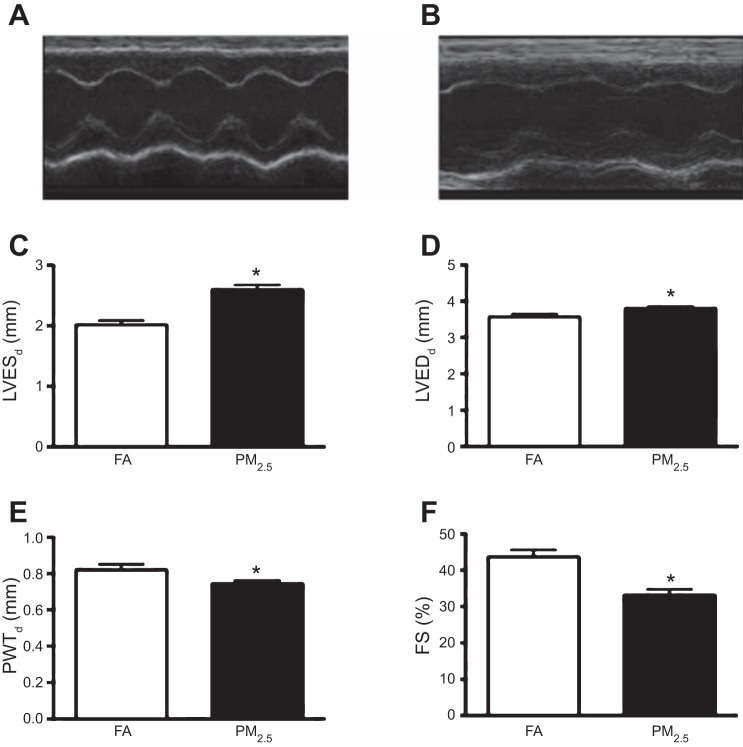
Fig. 1.
Transthoracic echocardiographic morphological and functional parameters in 3-mo-old FVB mice that were exposed to filtered air (FA) or particulate matter with diameter less than 2.5 μm (PM2.5) during perinatal development. Representative M-mode images obtained from FA-exposed mice (A) and PM2.5-exposed mice (B). C: left ventricular end-systolic diameter (LVESd). D: LV end-diastolic diameter (LVEDd). E: posterior wall thickness at systole (PWTs), from n = 10 mice in each treatment group and no more than 2 mice per litter. F: fractional shortening (FS). 5 beat cycles were captured, and 3 loops were averaged per assessment. *P < 0.05 was considered statistically significant.

