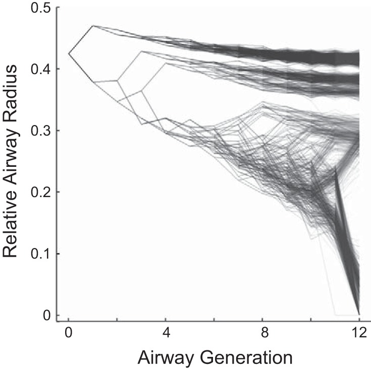
Fig. 2.

Example of self-organized bronchoconstriction used to explore pendelluft in a realistic context. Note that the constricted airways group together, leading to regionally clustered ventilation defects, and that there are substantial differences in constriction among daughter airways. The airways are shown as points and the connectivity among the airways as lines; darker areas illustrate higher local density.
