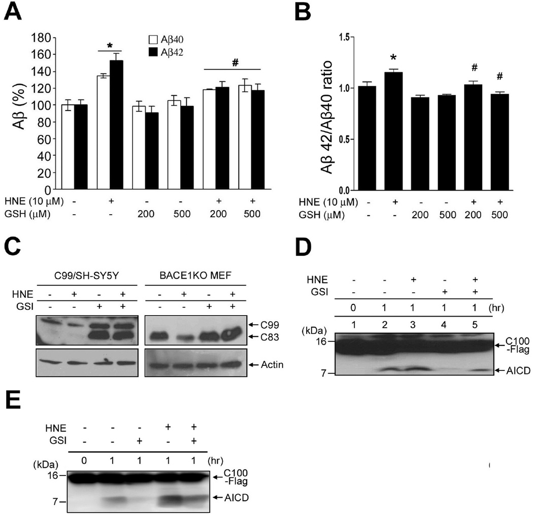
Figure 2.
The membrane lipid peroxidation product HNE increases Aβ42/Aβ40 ratio and AICD production. (A and B) HNE treating increased the amounts of secreted Aβ40, Aβ42 and Aβ42/Aβ40. After treating SH-SY5Y cells stably expressing mutant (Swedish) APP with HNE for 24 h, media were harvested and analyzed by sandwich ELISA for secreted Aβ40 (dark bars) and Aβ42 (light bars). In control cultures, the concentrations of Aβ40 and Aβ42 were 2067 ± 134 pg/ml protein and 848 ± 56 pg/ml, respectively (mean ± SD; n=3). *p < 0.01 vs. controls, #p < 0.01 vs. HNE-treated samples. (C) C99/SH-SY5Y cells (left panel) and BACE1KO MEF (right panel) were treated for 3 h with HNE (10 µM) in the presence or absence of the γ-secretase inhibitor DAPT (GSI; 1 µM). C99 and C83 levels were then analyzed using anti-APP-CTF antibody. (D and E) C100-Flag was incubated with the CHAPSO (D) or dodecyl-maltoside (E) solubilized lysate of SH-SY5Y cells at 37 °C; and the reaction was terminated at the indicated times by placing reaction tubes on ice. Reaction mixtures were separated in a 16% Tricine gel and subjected to immunoblotting for AICD using APP-CTF antibody.

