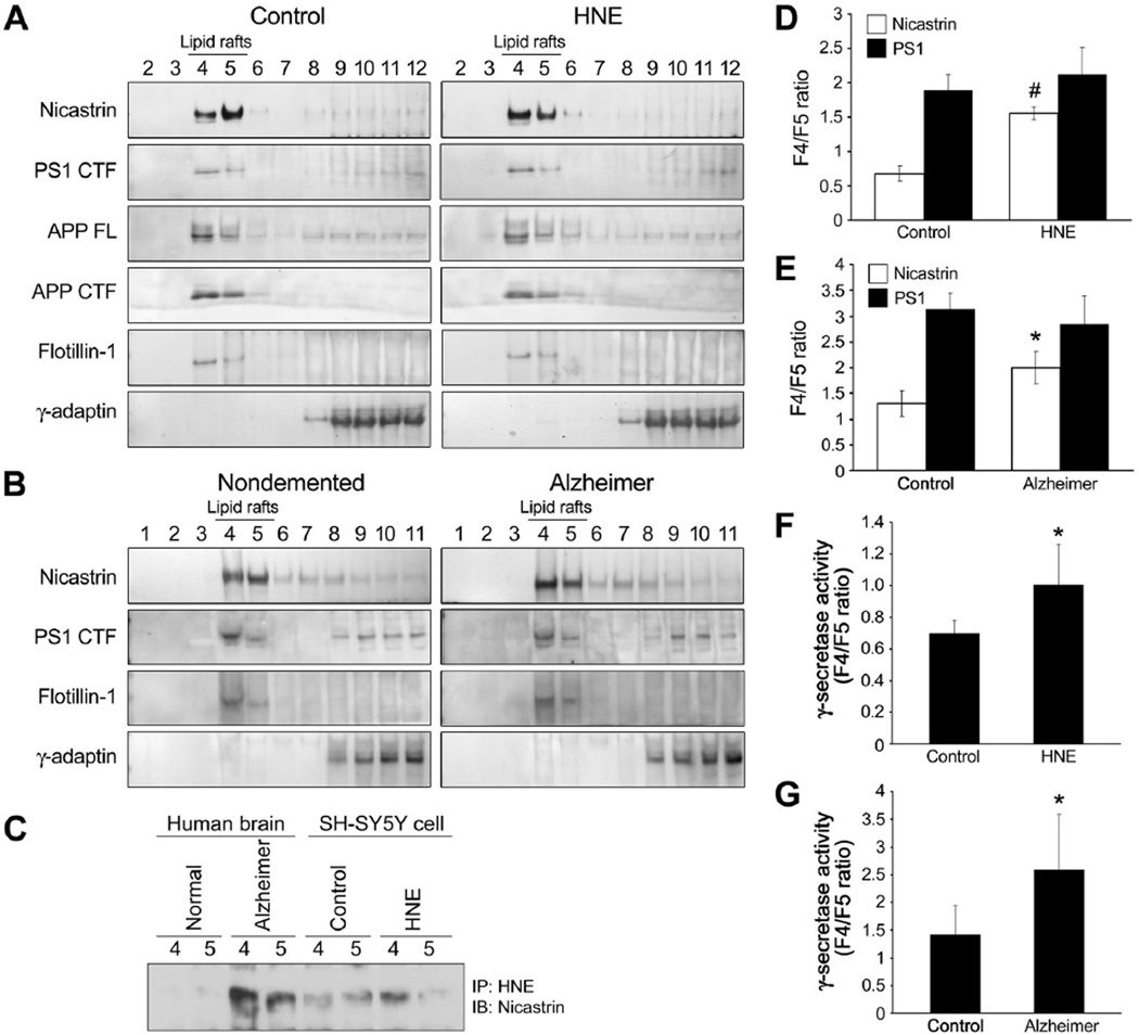
Figure 5.
Nicastrin is redistributed to an APP-enriched lipid raft fraction in AD brains and in response to direct exposure of neural cells to HNE. (A and B) Control and HNE-treated (10 µM for 3 h) SH-SY5Y cells (A), and AD and control inferior parietal lobes (B) were lysed in sodium carbonate buffer and subjected to flotation sucrose gradient centrifugation to isolate lipid rafts. Equal volumes of fractions were immunoblotted with antibodies against PS1 CTF, nicastrin, APP, APP CTF, flotillin-1 (a lipid raft marker), and γ-adaptin (a marker of clathrin-coated, non-raft membranes). (C) Lipid raft fractions (fractions 4 and 5) were immunoprecipitated using an antibody against HNE-modified proteins, and precipitated proteins were immunoblotted using anti-nicastrin antibody. (D and E) The signal intensities of PS1 CTF and nicastrin were quantified and fraction 4 versus fraction 5 (F4/F5) ratios were calculated for these proteins. The values shown are mean and S.D. (n=5; #p<0.01, *p<0.05 vs. controls). (F and G) γ-secretase activities in fractions 4 and 5, are reported as F4/F5 ratios. Values are the mean and S.D (n=5; *p<0.05 compared to control).

