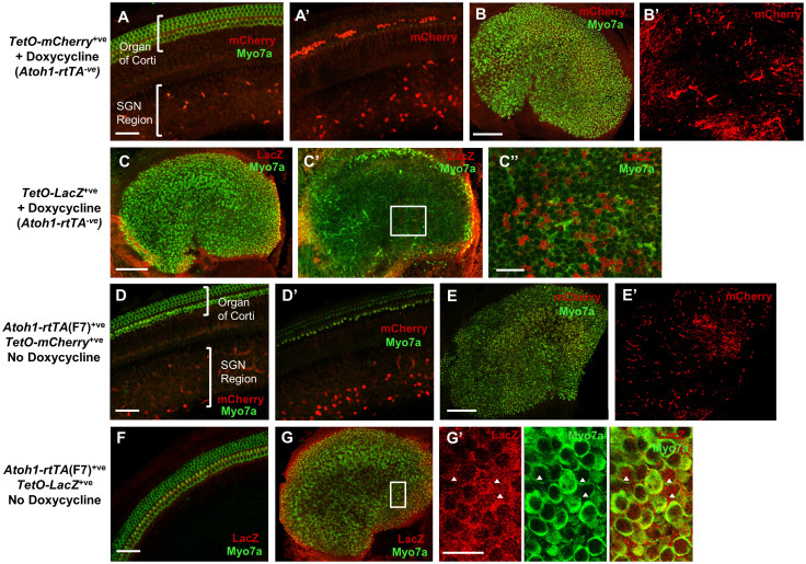
Figure 2. Basal activity of TetO-reporters and Atoh1-rtTA(F7) activity in the absence of doxycycline.
Representative confocal images of a TetO-mCherry+ve (Atoh1-rtTA−ve) control cochlea (A–A′) and utricle (B–B′) at P3 from mice that were administered doxycycline to test for basal expression of the TetO-mCherry reporter. mCherry+ve cells were detected in the SGN region of the cochlea (A–A′) and in the utricular stroma (B–B′). Note in A′ the labeled cells in the organ of Corti are red blood cells. No mCherry+ve cells were detected in the sensory region of either the cochlea or utricle. (C–C′) Representative confocal images of a TetO-LacZ+ve (Atoh1-rtTA−ve) control utricle at P3 from mice that were administered doxycycline to test for basal expression of the TetO-LacZ reporter. Some LacZ+ve SCs but no LacZ+ve HCs were detected. (C″) High magnification images of the region outlined by the white square in C′. Representative confocal images of an un-induced Atoh1-rtTA(F7)+ve; TetO-mCherry+ve control cochlea (D–D′) and utricle (E–E′) at P3 that did not receive doxycycline. mCherry+ve (red) cells were detected in the SGN region of the basal turn of the cochlea (D–D′) and in the urticular stroma (E–E′). No mCherry+ve cells were detected in the sensory region of either the cochlea or utricle. Representative confocal image of an un-induced Atoh1-rtTA(F7)+ve; TetO-LacZ+ve control cochlea (F) and utricle (G–G′) at P3 that did not receive doxycycline. Several HCs (myo7a+ve, green) expressed LacZ (red) in the medial region of the utricle (G–G′) and no LacZ+ve cells were detected in the cochlea (F). (G′) High magnification image of the region outlined by the white square in (G). Arrowheads denote LacZ+ve HCs. Myo7a (green) was used to label HCs in most panels. Scale bar: 50 μm in (A–A′), (D–D′), and (F), 100 μm in (B–C′), (E), and (G), and 20 μm in (C″) and (G′).

