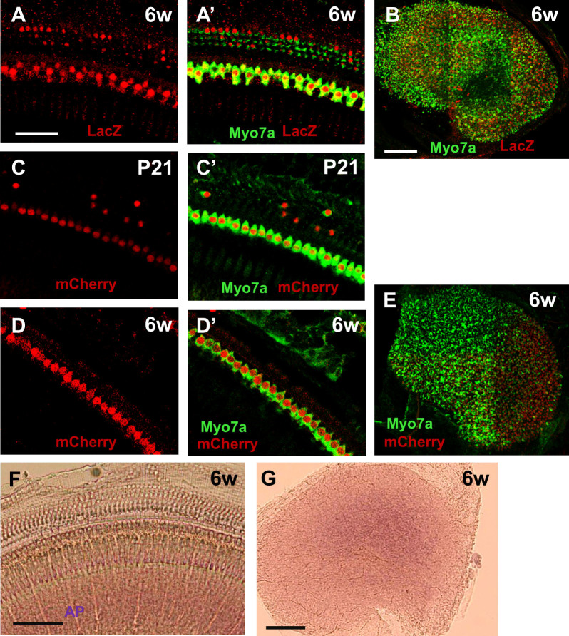
Figure 6. TetO-reporter expression was still present at 6 weeks of age.
Representative confocal images of LacZ+ve (red) cells in the middle cochlear turn (A) and utricle (B) of Atoh1-rtTA(F7)+ve; TetO-LacZ+ve mice at 6 weeks of age after doxycycline treatment ended at P3. (A’) is a merged image with HCs labeled by myosin VIIa (Myo7a, green). Representative confocal images of mCherry+ve (red) cells in the middle cochlear turn of Atoh1-rtTA(F7)+ve; TetO-mCherry+ve mice at P21 (C) and 6 weeks of age (D) and in the utricle at 6 weeks of age (E) after doxycycline treatment ended at P3. (C’, D’) are merged images with HCs labeled by myosin VIIa (Myo7a, green). Representative alkaline phosphatase (AP) staining images at 6 weeks of age in the cochlea (F) and utricle (G) of Atoh1-rtTA mice from the F7 lineage. Scale bars: 50 μm in (A–A′), (C–D) and 100 μm in (B, E–G).

