. 2014 Mar 5;2(5):634–650. doi: 10.1039/c3bm60319a

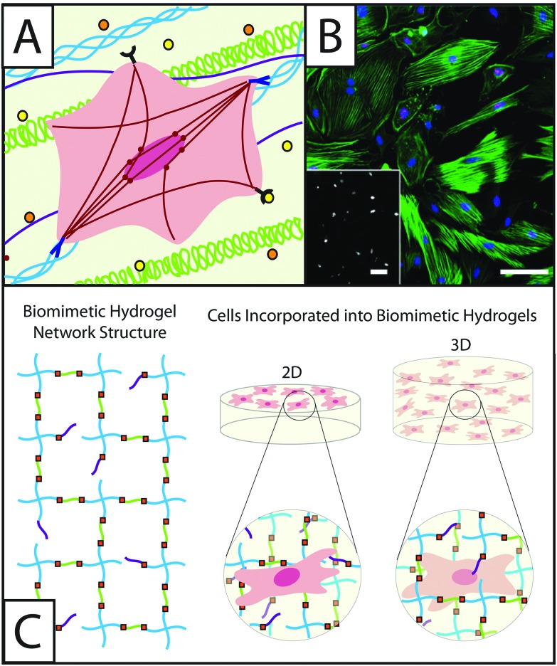
Fig. 1. Understanding the role of fibroblasts in healing and disease using hydrogel-based culture models. (A) During normal or misregulated healing in tissues throughout the body, fibroblasts are presented with a variety of signals that affect their function and activation into wound-healing cells, myofibroblasts. These extracellular cues include cytokines, such as TGFβ1 or PDGF (orange and yellow circles), that are released into the ECM by other fibroblasts, immune response cells, and epithelial cells, as well as extracellular matrix proteins, such as collagen (blue triple helix), hyaluronic acid (green), and fibronectin (purple). Interactions between fibroblasts and the temporally changing matrix lead to cytoskeletal reorganization, including expression of α-smooth muscle actin (αSMA) stress fibers (red), as well as alterations in gene expression and secretion of enzymes and proteins for matrix remodeling. The presence of organized αSMA fibers is often used to identify contractile myofibroblasts.5 (B) Traditionally fibroblasts (here, primary rat lung fibroblasts) have been cultured on tissue culture polystyrene, a stiff substrate known to induce activation, as shown here by cells (blue nuclei) that stain positive for αSMA (green stress fibers) and a proliferation marker (red Ki67). Adapted from reference 7. (C) Hydrogel-based culture models afford varying degrees of control of matrix biophysical and biochemical properties to understand their role in healing and disease. Here, a hydrogel with an ideal network structure is depicted with a 4-arm monomer linked by small peptides (green), which can be functionalized to be cell adhesive or proteinase degradable, as well as pendant biofunctional groups (purple). Fibroblasts have been cultured on and within hydrogel scaffolds to understand how individual or combinations of extracellular cues influence their function and fate in healing and disease.

Fig. 1