. 2014 Mar 5;2(5):634–650. doi: 10.1039/c3bm60319a

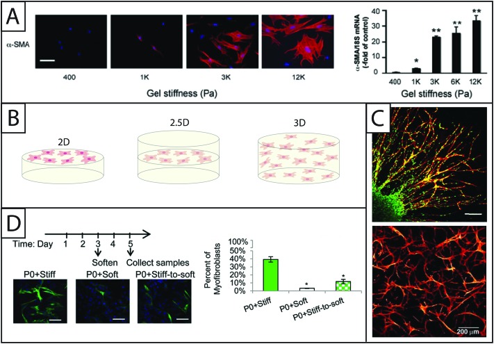
Fig. 5. Increasing dimensionality: Examining fibroblast response within static and dynamic model systems from two to three dimensions. (A) Polyacrylamide gels of various moduli modified with collagen I have been a dependable system for examining the effect of modulus on fibroblasts in 2D culture. Increasing the modulus of the hydrogels increases expression of αSMA (here, rat hepatic fibroblasts, αSMA immunostaining (left) and qRT-PCR (right)).70 This result supports the growing body of evidence that substrate stiffness has a significant impact on fibroblast activation, corresponding with in vivo observations of myofibroblast persistence with increased tissue stiffening. Adapted from reference 70. (B) While many seminal studies have been made in 2D culture, cells natively are surrounded by matrix. Towards addressing this, fibroblasts have been cultured in 2D, 2.5D, and 3D geometries to understand the effect of polarization on cell behavior. 2D cell culture geometry, where fibroblasts are seeded on top of hydrogels, has been commonly used to study fibroblasts in vitro (left). 2.5D cell culture geometry is an emerging technique that allows for a decrease in polarization while maintaining the ability to image cells, a key to assessing αSMA stress fiber formation and cell activation (middle). 3D cell culture geometry is used to accurately mimic the lack of cell polarization within the in vivo environment (right); however, cells must be able to degrade and spread in the matrix of interest. (C) For example, in 3D culture, human foreskin fibroblasts (top) have been cultured within PEG gels that were rendered degradable by the incorporation of MMP-degradable peptide sequences. Fibroblasts encapsulated within a fibrin clot were shown invading the synthetic hydrogel by degrading and migrating through the network (scale bar, 150 μm). Human foreskin fibroblasts directly encapsulated in these PEG hydrogels (bottom) formed a network with single cells migrating considerably within the gel over 30 days.76,79 Adapted from references 76 and 79. (D) Towards capturing temporal changes in the cell microenvironment, in vitro culture systems whose properties can be changed in time utilizing external triggers have been created. For example, photodegradable hydrogels have been utilized to trigger a temporal decrease in matrix modulus with light. Here, when the modulus of the hydrogel was reduced after three days of cell culture, fewer valve fibroblasts (VICs) expressed αSMA stress fibers (green) compared to VICs continually cultured on stiff hydrogels; however, the number of αSMA positive cells was still higher than VICs continually cultured on soft substrates, indicating that some portion of the fibroblasts can be de-activated with changes in matrix modulus at this time point in culture.19 Adapted from reference 19.

Fig. 5