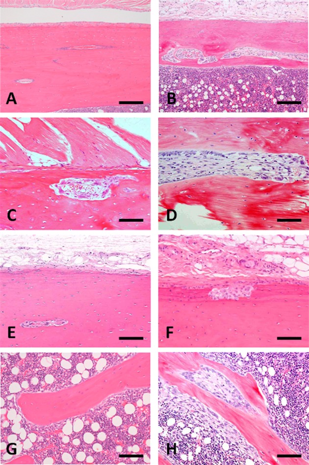
Fig. 5.

Photomicrograph of femoral bone changes in non-treated control marmosets at weeks 13 (A and G) and 26 (E) and the 5/6Nx marmosets at weeks 13 (B, C, D and H) and 26 (F). Histological structure of femoral cortical bone at week 13(A) and 26 (E) and trabecular bone at week 13 (G) in non-treated control animals. Bone resorptive change (B and C) and microfibrosis (D) in the lumens of Haversian canals of cortical bone and a decrease in trabecular bone with surrounding fibrous tissue (H) present in 5/6Nx animals at week 13. Resorptive changes appear in the subperiosteal cortical bone (F) of 5/6Nx animals at week 26. Animal Nos.: A and G, 005; B, C, D and H, 107; E, 007; F, 113. H-E stain. Scale bar = 100 μm
