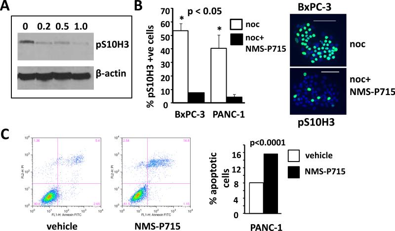
Figure 2. NMS-P715 treated PDAC cell lines exhibit bypass of the spindle assembly checkpoint and apoptosis.
A. Western analysis using a pS10H3 antibody of human PANC-1 cells treated with either NMS-P715 (μmol/L) or vehicle (0) as indicated for 72h. B. Left panel: Cells were treated with nocodazole (noc) or noc plus NMS-P715 (noc+NMS-P715). Percentage pS10H3 positive cells in each treatment group is indicated in the graph. Right panel: representative image of BxPC-3 cells treated either with noc or noc + NMS-P715. pS10H3 positive cells are green. Nuclei (blue) are stained with DAPI. Scale bar =100 μm. C. Flow cytometric analysis of Annexin V (Annexin-FITC) and propidium iodide (PI) labeled PANC-1 cells treated with 1 μmol/L NMS-P715 for 40h. Histogram shows the percentage of apoptotic cells calculated by combining the fraction of Annexin V positive (bottom right) and double positive (top right) cells. Significance was measured using a t-test (B) or χ2 test (C).

