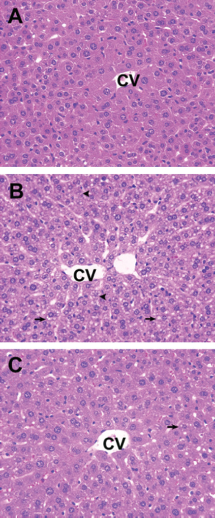
Fig. 3.
Histopathological changes in the liver of different groups. (A) Control mice. (B) Ethanol (EtOH) treatment induced prominent microvesicular steatosis (arrows) along with necrosis (arrowheads) in the liver. The necrotic hepatocytes are characterized by cell enlargement and nuclear dissolution. (C) Livers from silymarin supplemental group showed only microvesicular steatosis, which was less extensive than in livers from mice receiving EtOH alone. CV, central vein. ×40.

