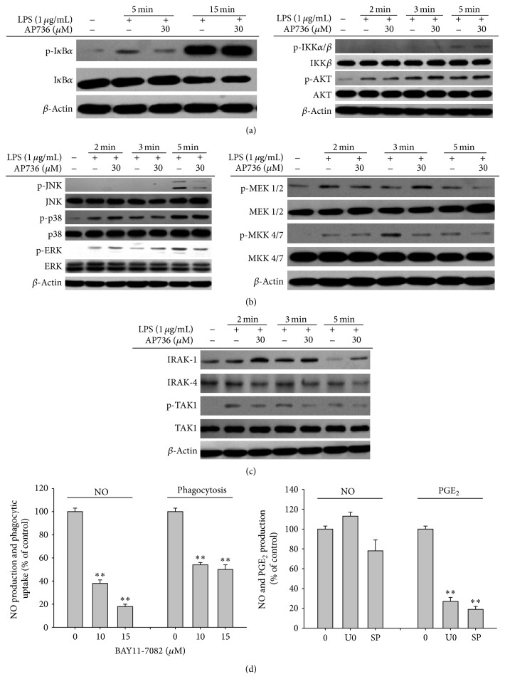
Figure 5.
Effects of AP736 on signalling cascades upstream of NF-κB and AP-1 activation. ((a), (b), and (c)) Phosphoprotein or total protein levels of IκBα, IKK, AKT, p38, ERK, JNK, MEK1/2, MKK4/7, TAK1, IRAK1, IRAK4, and β-actin were determined by immunoblot analysis of cell lysates using phosphospecific or total protein antibodies. (d (left panel)) RAW264.7 cells (1 × 10⁶ cells/mL) were treated with FITC-dextran (1 mg/mL) or LPS (1 μg/mL) in either the presence or absence of BAY 11-7082 (10 and 15 μM). Cells were then incubated for 30 min (phagocytic uptake assay) or 24 h (NO assay). The levels of NO in culture supernatants were determined using the Griess assay. (d (right panel)) RAW264.7 cells (1 × 10⁶ cells/mL) were treated with LPS (1 μg/mL) in the presence or absence of SP600125 (SP, 25 μM) or U0126 (U0, 25 μM). Cells were incubated for 6 h (PGE2 assay) or 24 h (NO assay). The levels of NO or PGE2 in culture supernatants were determined using the Griess assay and EIAs, respectively.

