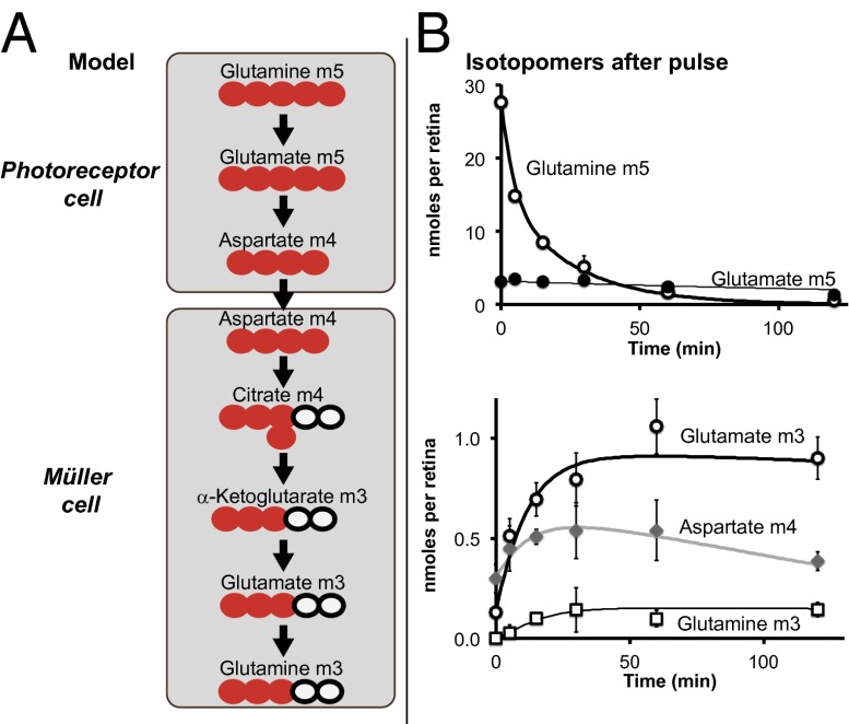
Fig. 5.
Pulse–chase analysis of U-13C Gln in retina. (A) Schematic model for the role of Asp as a carrier of oxidizing power between retinal neurons and glia. Red circles represent the 13C carbons, and black circles represent the 12C carbons. (B) 13C labeling of Asp, Glu, and Gln from the pulse of U-13C Gln. (Upper) The M5 Gln and Glu are derived directly from the pulse of 5 mM U-13C Gln. After 5 min, the medium was changed to 5 mM unlabeled Lac with no added Gln. Unlabeled Gln was not included in the chase because the intense signal from the added Gln would have obscured the isotopomer signals we intended to quantify. The retinas were subsequently harvested at the indicated times after the pulse. (Lower) The M4 Asp derived from oxidation of Glu via the TCA. The M3 Glu is made by further oxidation via citrate, and M3 Gln is made only in MCs by Gln synthetase. (n = 6).

