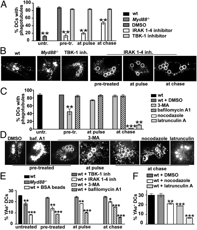
Fig. 2.
Phagosome tubulation requires TLR4-MyD88 signaling and an intact cytoskeleton and has a moderate impact on MHC-II-antigen presentation. BMDCs from WT or Myd88−/− mice were untreated or pretreated, treated at pulse or at chase with vehicle (DMSO) or inhibitors as indicated, and pulsed with LPS/OVA-TxR beads, Eα beads, or LPS/BSA beads. (A–D) BMDCs pulsed with LPS/OVA-TxR beads and treated as indicated were analyzed by live cell imaging after a 2.5-h chase. (A and C) The percentage of DCs that showed phagotubules during 2 min of analysis was calculated for 10 cells per experiment, five independent experiments. (B and D) Frames from representative movies of cells treated as indicated. (E and F) Cells treated as indicated, pulsed with Eα beads (or LPS/BSA beads as a control), and chased for 4 h were labeled for surface MHC-II:Eα52–68 by YAe antibody and analyzed by flow cytometry. Shown are the percentages of positive cells. Error bars represent mean ± SD.

