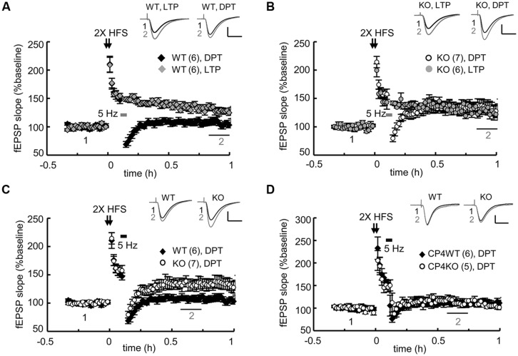
FIGURE 3.
Genetic ablation of CPEB3 impaired depotentiation in the SC-CA1 pathway of hippocampal slices. The slices prepared from CPEB3 (A) WT and (B) KO mice were stimulated with two trains (2X) of HFS to induce LTP. To erase the 2X HFS-evoked LTP, 3 min of 5-Hz tetanus was applied 5 min after the HFS to induce DPT. Only DPT but not LTP was defective in the KO slices (WT, LTP: 128.80 ± 5.12%, DPT: 106.46 ± 5.28%, P < 0.01 at 50–60 min; KO, LTP: 127.04 ± 8.61%; DPT: 134.12 ± 10.57%, P = 0.26 at 50–60 min). (C) Depotentiation was impaired in the adult CPEB3KO mice (WT: 107.72 ± 4.71%, KO: 133.36 ± 9.35%, P < 0.01 at 30–40 min after stimulation). (D) Deletion of cpeb4 gene did not affect DPT (CP4WT: 112.22 ± 6.63%; CP4KO: 115.87 ± 7.45%, P= 0.20 at 30–40 min after stimulation). The numbers in parentheses represent the numbers of recorded slices isolated from 5 to 6 male mice. All of the data are expressed as the mean ± SEM. The statistics were performed with Student’s t-tests. The traces represent the baseline (black line, 1) and the indicated time after stimulation (gray line, 2). Calibration: 0.5 mV, 20 ms.

