FIGURE 6.
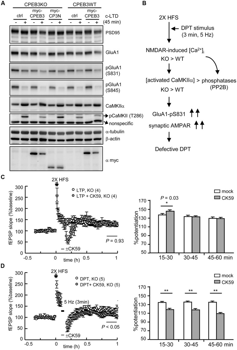
The defective form of DPT in the CPEB3-deficient hippocampus was rescued by the transient inhibition of CaMKIIα activity. (A) The neurons infected with or without the lentivirus expressing myc-CPEB3 or the N-terminus of CPEB3 (myc-CP3N) were stimulated with or without a 3-min pulse of 20 μM NMDA and then harvested 45 min later for Western blotting. (B) The results from c-LTD-treated KO neurons suggested that the DPT defect in the KO hippocampal slices might have been caused by elevated NMDAR-mediated calcium influx, which shifted the balances between calcium/calmodulin-dependent kinase and phosphatase, i.e., CaMKIIα and calcineurin (PP2B), respectively. Consequently, the increased Ser831 phosphorylation of GluA1 failed to decrease the synaptic AMPAR level to induce DPT. (C) The 2X HFS-induced LTP in the KO slices was not affected by the 3-min application of the CaMKIIα inhibitor CK59 (10 μM; mock: 130.14 ± 7.81%, CK59: 130.41 ± 6.41%, P = 0.93 at 50–60 min). (D) The application of CK59 during the 3-min of 5-Hz stimulation facilitated DPT in the KO slices (mock: 133.44 ± 8.47%, CK59: 111.22 ± 6.47%, P < 0.01 at 50–60 min after stimulation). The histograms display the % potentiation during three time frames (15–30, 30–45, and 45–60 min). The numbers in parentheses represent the numbers of recorded slices isolated from 3 to 5 male mice. All of the data are expressed as the mean ± SEM. The statistics in (C) and (D) were performed with Student’s t-tests. Single and double asterisks represent P < 0.05 and P < 0.01, respectively.

