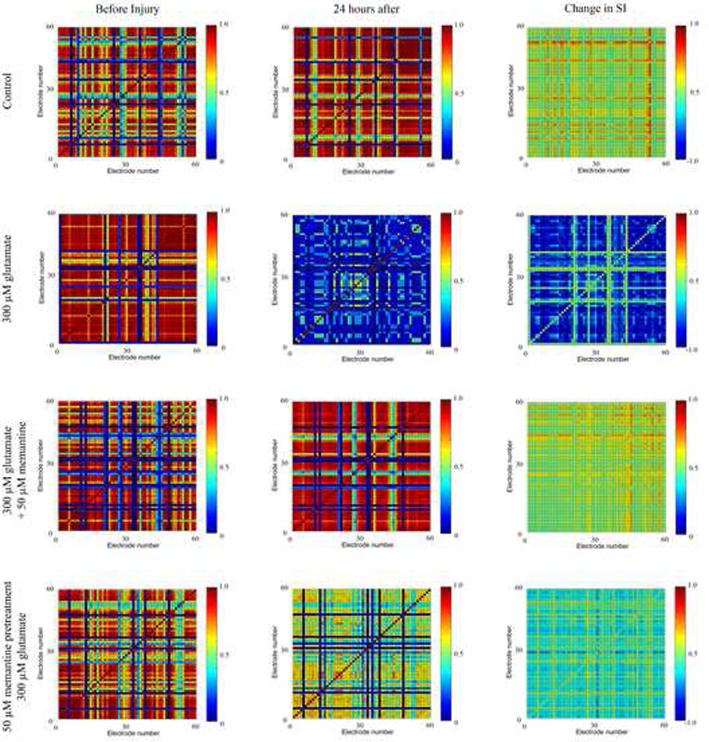
Figure 1. The ability of memantine to protect the synchronized firing between electrode pairs.
A representative MEA for each condition is shown. SI=synchronization index. Column 1 shows the synchronization grids before drug treatment, column 2 shows the synchronization grids 24 hours after drug treatment, and column 3 shows the change in synchronization (CS) as a result of drug treatment. In the left and middle columns, a red color designates that the two electrodes have a high SI, indicating that both electrodes frequently record a spike within the same burst. A dark blue color designates little to no synchronization between the neurons whose action potentials are recorded on those electrodes. In the third column, a red or yellow color indicates an increase in the SI (positive CS), a pale green color represents no change in the SI, and a blue color represents a decrease in the SI (negative CS).

