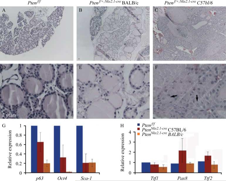
Figure 6.
Hematoxylin and eosin staining of (A) control and (B and C) heterozygous thyroids at 2 years of age. (A) Control thyroids show normal histology with structured colloid-filled follicles. (B) Heterozygous Ptenflox/C;Nkx2.1-cre BALB/c thyroids show enlarged follicles in the middle of areas of atrophy. (C) Heterozygous Ptenflox/C;Nkx2.1-cre thyroids in C57BL/6 background display thyroid structure that was altered, with normal areas presenting not only with colloid-filled follicles but also with focal hyperplasia, small nonencapsulated areas of hypercellularity with solid and/or microfollicular patterns. (D–F) Corresponding high magnification pictures. Arrows in (F) indicate cells with more than one nucleus. (G and H) Real-time PCR analysis of key stem cell genes. (G) Expression levels of p63, Oct4, and Sca-1 showing the decrease of all stem cell markers in both C57BL/6 and BALB/c homozygous mutants compared with control thyroids. (H) Thyroids in the C57BL/6 background display increased expression of the transcriptional factors Pax8 and Ttf2. Such an increase was not detected in the BALB/c homozygous mutant thyroids.

