
Figure 1.
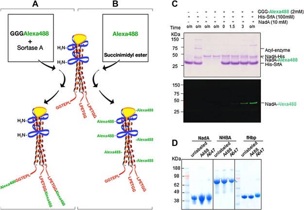
Sortagging and amine-labeling of NadA. (A) Model of the three-dimensional organization of NadA with dimeric and trimeric coiled-coil-rich stalk regions (blue, red) and globular head (yellow). (A) Sortagging (A) inserts fluorochrome molecules in a site-specific manner to the C terminus of the elongated coiled-coil-rich stalk region while (B) chemical labeling inserts flurochrome molecules at random to solvent accessible amine groups. (C) C-terminal NadA labeling using SrtAStaph. NadA with a C-terminal LPETGG followed by HA and His tag (Nad-His) (10 µM) was incubated with 100 µM SrtA with and without GGG-Alexa 488 (2 mM). The reaction was terminated at various times with Laemmli sample buffer, subjected to SDS–PAGE, and analyzed by Coomassie staining (upper panel) and fluorescent gel scanning (bottom panel). (D) Integrity of amine-labeled MenB vaccine antigens analyzed by SDS–PAGE and Coomassie staining.