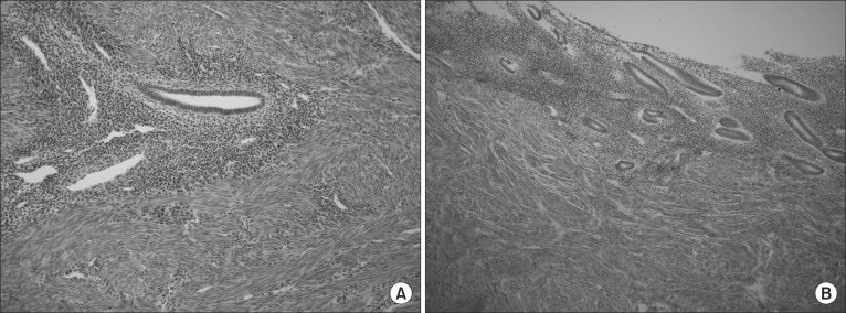
Fig. 4.
Histologic findings from the resected specimen. (A) Uterine myometrium (H&E, × 200). Microscopically, the uterus shows typical adenomyosis features, such as multifocal atrophic endometrial glands and stroma in the background of proliferating smooth muscle cell bundles. (B) Uterine endometrium (H&E, × 100). The atrophoic endometrium (< 1 mm thickness) is seen in the lower segment of the uterus.

