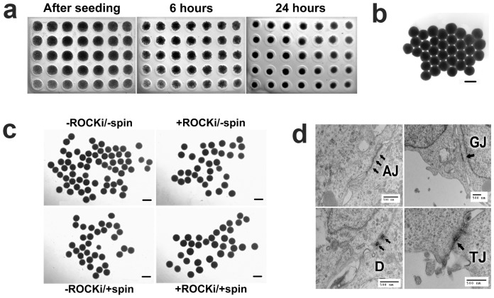
Figure 1. BG01V/hOG cells formed hEBs in the microwells at a cell seeding density of 15,000 hESCs per microwell.
(a) Condensation of the hESC suspension within the microwells was progressive and evident after 6 hrs of incubation. (b) hEBs were compact and spherical and be able to be extracted intact from the microwells after 24 hrs of incubation. (c) The freshly extracted hEBs formed under four different conditions (ROCKi: ROCK inhibitor; Spin: centrifugation). (d) Internal structural organization among the cells within the freshly extracted hEBs (formed under -ROCK/-spin condition) was demonstrated by the presences of cell-cell junctions, i.e., adherence junctions (AJ), gap junctions (GJ), desmosomes (D), and tight junctions (TJ), in TEM images. All treatments were performed with aliquots from the same cell suspension. Microwell diameter 820 µm. Scale bars 500 µm.

