
Figure 2. H9 hESCs formed hEBs in the microwells at a cell seeding density of 25,000 hESCs per microwell.
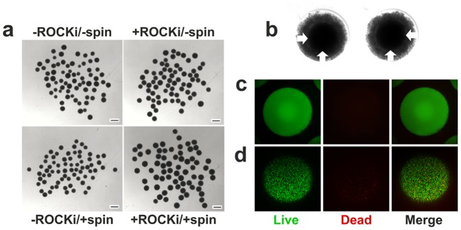
(a) Freshly extracted hEBs formed under four different conditions in microwells after 24 hrs of incubation. (b) A closer look at hEBs formed under -ROCK/-spin condition after 24 hrs of incubation before extraction indicated the presences of a compact core (arrows) and a loosely-aggregated corona. (c) Live-dead staining of the formed hEBs under -ROCK/-spin condition after 24 hrs of incubation before transfer to suspension culture indicated high viability (>90%) of hESCs both at the core and the corona (SYTO 10 green-fluorescent nucleic acid stain for all cells; DEAD Red (ethidium homodimer-2) nucleic acid stain for dead cells). (d) Confocal microscopic images of live/dead staining of freshly extracted hEBs (formed under -ROCK/-spin condition) indicated high viability (>85%) of the hESCs. Note that cells at the corona of the hEBs were sloughed off during the transfer process. Scale bars 500 µm.