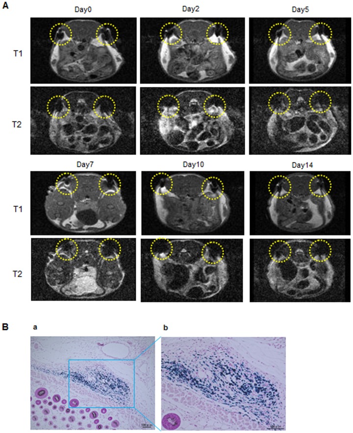
Figure 7. MR imaging of ASCs labeled with TMADM-03 under the skin and Prussian blue staining.
A: In vivo MR imaging of ASCs (2×10⁶) labeled with TMADM-03 (30 µg-Fe/mL) under the skin in a cross-section figure from the head of the mouse for 14 days. The two yellow dotted circles show the transplanted ASCs labeled with 30 µg-Fe/mL of TMADM-03. These images were obtained using a 1T MRI instrument (MR Technology). B: Prussian blue staining of the transplanted ASCs labeled with TMADM-03.

