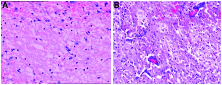
Figure 1.
Hematoxylin and eosin staining (magnification, ×100). (A) Day 1 of patients following minimally invasive evacuation of hematoma. The perihematoma tissues were infiltrated by inflammatory cells and appeared to be loose. The extravascular space was expanded. (B) Day 1 of patients following mild hypothermia and minimally invasive evacuation of hematoma. Vascular congestion was observed in the tissues surrounding the hematoma in the acute phase. The size of nerve cells was reduced, karyopyknosis was observed, Nissl substance had disappeared and a large number of neutrophils and lymphocytes had infiltrated.

