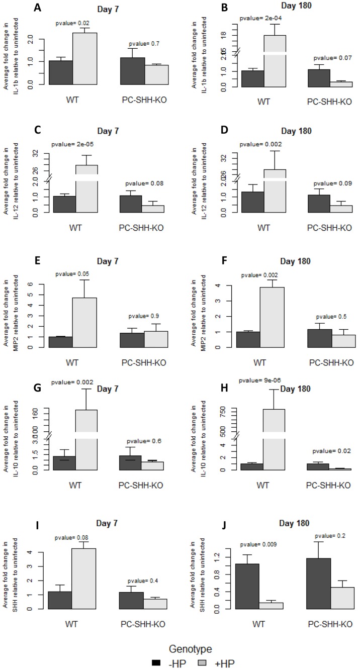
Figure 3. Effect of H. pylori on SHH and cytokines' expression in WT and PC-SHHKO mouse stomachs, day 7 and day 180 post-inoculation.
RNA was extracted from stomachs of uninfected and H. pylori-infected wild type (WT) and parietal cell-specific SHH knock out (PC-SHH-KO) mice 7 and 180 days post-inoculation. Expression of genes was measured by qPCR and two-way ANOVA test was performed, followed by Bonferroni test to compare uninfected (-HP) with H. pylori infected group (+HP) in each genotype. The graphs show average fold change in expression of IL-1β (A, B), IL-12 (C, D), MIP-2 (E, F), IL-10 (G, H) and SHH (I, J) upon H. pylori infection relative to uninfected condition. Bars represent the mean ± SEM, n = 3-4 per group.

