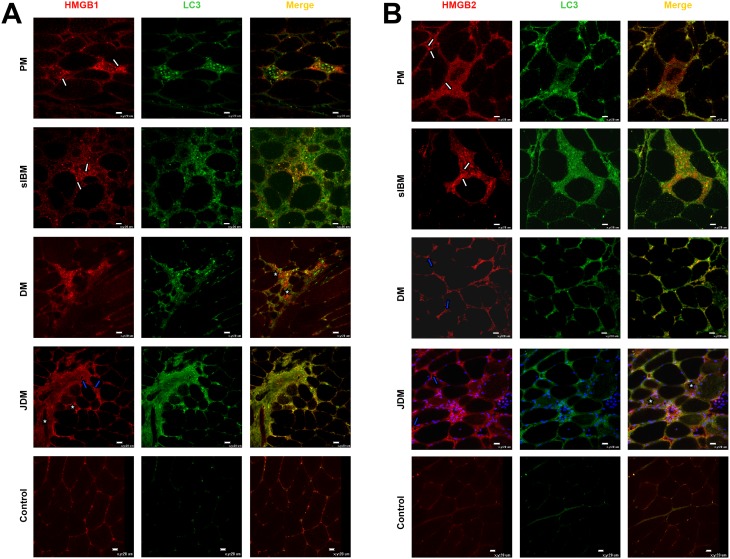
Figure 3. Representative micrographs showing immunolocalization of alarmins (A) HMGB1 and (B) HMGB2 in IIM and controls.
Both HMGB1 and HMGB2 are highly expressed in all IIM samples, mainly in association with immune infiltrates (white arrows), blood vessels (blue arrows), nuclei of myofibers, and occasionally with muscle fiber cytoplasm (white asterisks). Both HMGB1 and 2 co-localize with LC3 in association with muscle infiltrating cells and myofibers (white asterisks). In control muscle LC3 and HMGB1 and HMGB2 staining is weak or absent. Original magnification x40; bar: 20 µm.

