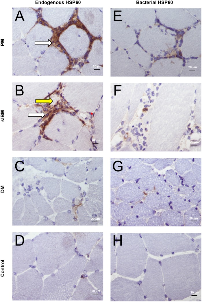
Figure 4. Endogenous and bacterial HSP60 in IIM and control muscles.
Endogenous HSP60 is highly expressed in all IIM (A–C), mainly in association with inflammatory infiltrates (white arrows), vascular endothelial cells (red asterisk), myofibers surrounded or invaded by immune infiltrates (yellow arrow). In control muscles only occasional capillaries were positive for endogenous HSP60 (D). Bacterial HSP60 was occasionally observed associated with muscle infiltrates in all IIM muscles (E–G), particularly PM (E) and sIBM (F), but not controls (H). Original magnification x40; bar: 20 µm.

